1. Primary Interventional Records & Logs
The following documents provide the administrative and procedural context for the emergency endovascular intervention. They outline the recorded timeline, technical parameters, and imaging‑related metadata associated with the procedure.
-
Handwritten Catheterization Lab Log (心カテ検査記録)
Covers the period from 22:35 (August 24) to 01:35 (August 25). Documents real‑time procedural milestones, vascular access attempts, and subsequent hemodynamic changes. -
Formal Coronary Summary Report (CORONARY REPORT)
Provides the institutional summary of angiographic findings, stent deployment details, and contrast‑flow characteristics. -
→ Master Video Track Chronology Matrix (PCI動画と時間の対応表)
Maps each fluoroscopic video file (XA0001–XA0090) to its corresponding intraoperative timestamp and associated coronary events.
2. Discrepancy Between Institutional Statements and Native Imaging Records
Official briefings provided by hospital representatives, as well as subsequent summaries issued by legal and law‑enforcement authorities, consistently state that
“the judicial autopsy identified no medical errors or procedural trauma.”
The native fluoroscopic imaging archived below presents findings that do not align with this conclusion.
Continuous frame‑by‑frame review of the angiographic tracks demonstrates multiple sites of vascular injury that are not compatible with spontaneous or natural disease processes.
These differences highlight a significant discrepancy between the institutional narrative and the objective procedural imagery.
1. Radiopaque Object of Uncertain Origin (XA0056.mp4)
XA0056.mp4:SHA256:XXX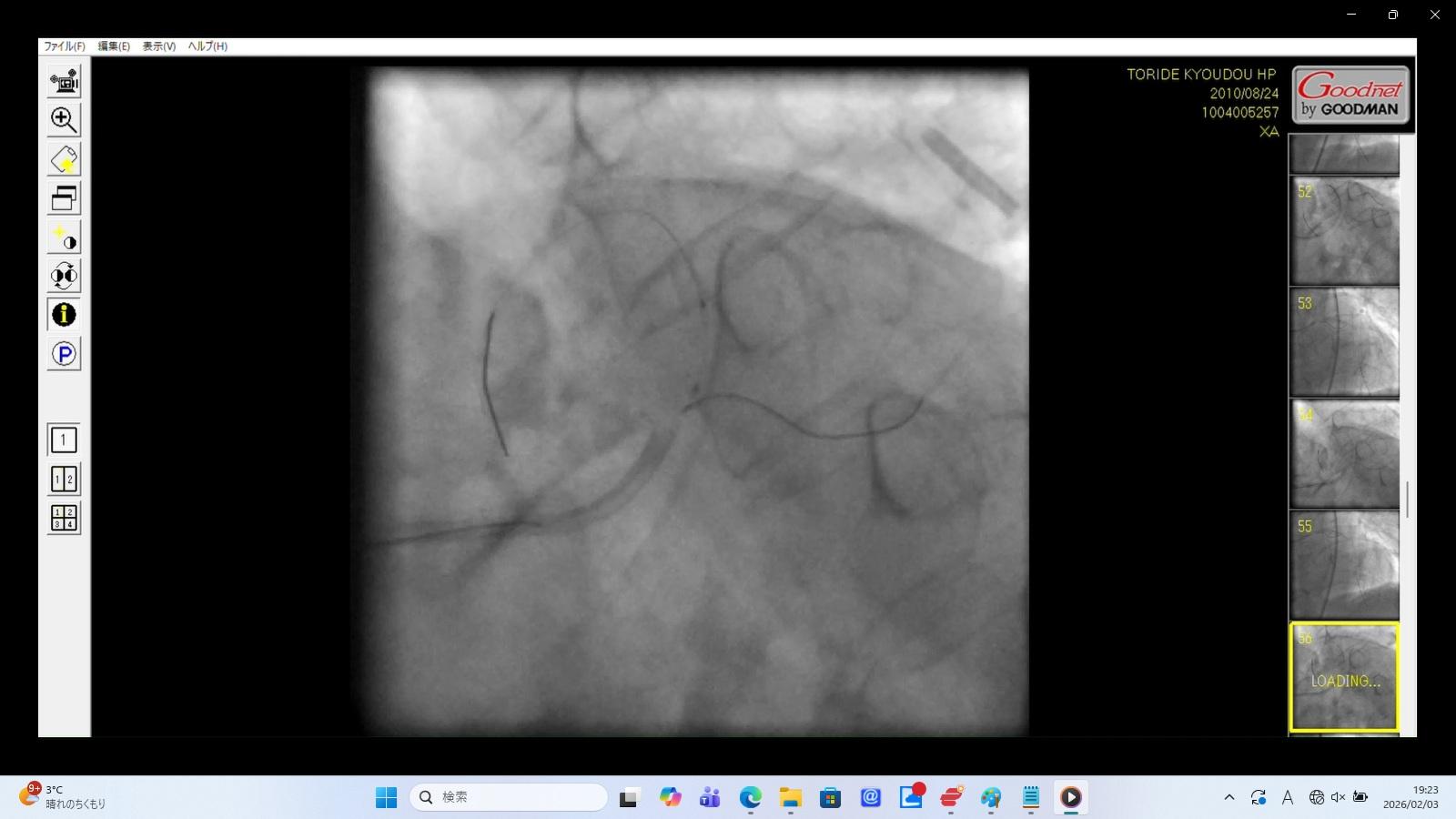
Medical Findings:
Radiopaque Cylindrical Structure:
A distinct cylindrical, metallic‑density object is visible in the upper‑right quadrant of the fluoroscopic frame.
Pulsation‑Synchronous Motion:
The object moves in coordination with cardiac pulsation, suggesting a location within the thoracic cavity or pericardial space.
Interpretive Note:
The object is not referenced in the available procedural records or autopsy documentation.
Its appearance is consistent with an unaccounted intrathoracic medical device fragment, such as a catheter or stent component.
2. Coronary Flow Irregularity Consistent with Dissection (XA0020.mp4)
XA0020.mp4:SHA256:XXX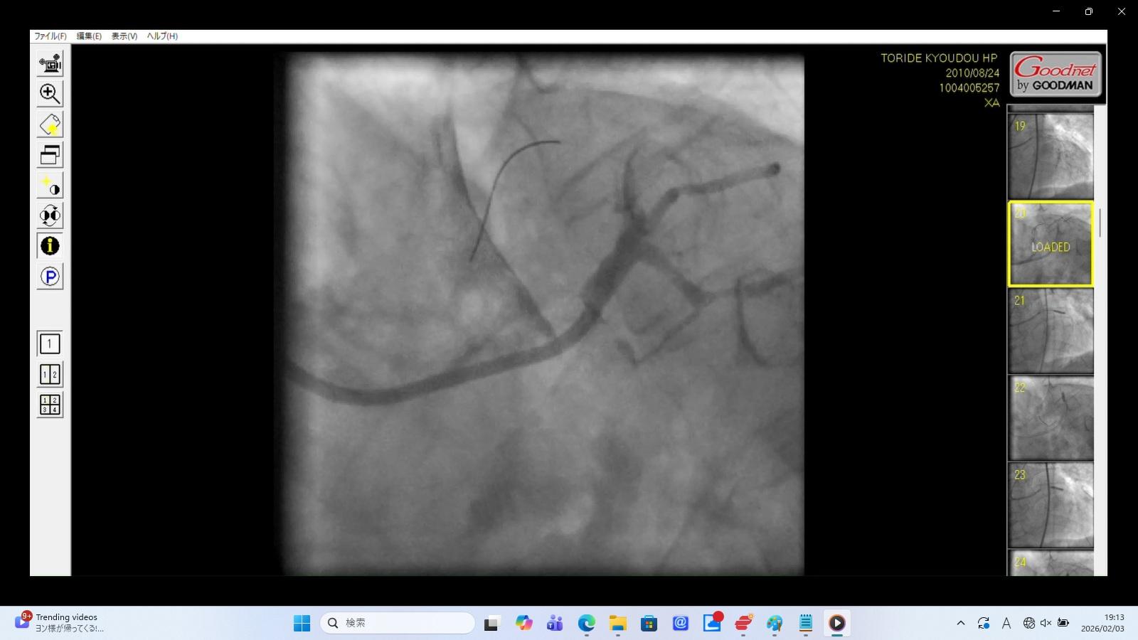
Finding:
Contrast media enters the LAD slowly and expands into a non‑anatomical plane, producing a pattern suggestive of contrast tracking outside the true lumen.
Interpretive Note:
The imaging characteristics are consistent with coronary artery dissection.
The catheter tip appears positioned within a false lumen, and the observed “slow flow” corresponds to contrast accumulating between vessel wall layers.
3. Left Main Trunk (LMT) Structural Injury (XA0034.mp4)
XA0034.mp4:SHA256:XXX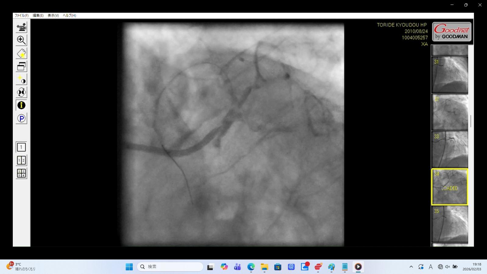
Finding:
A false lumen is visible within the Left Main Trunk (LMT), accompanied by contrast leakage beyond the vessel boundary.
Interpretive Note:
These findings indicate a significant structural injury to the LMT, involving both intramural dissection and extraluminal contrast extravasation.
Such patterns are consistent with major periprocedural vascular trauma.
4. LAD Perforation (XA0066.mp4, XA0072.mp4)
XA0066.mp4:SHA256:XXX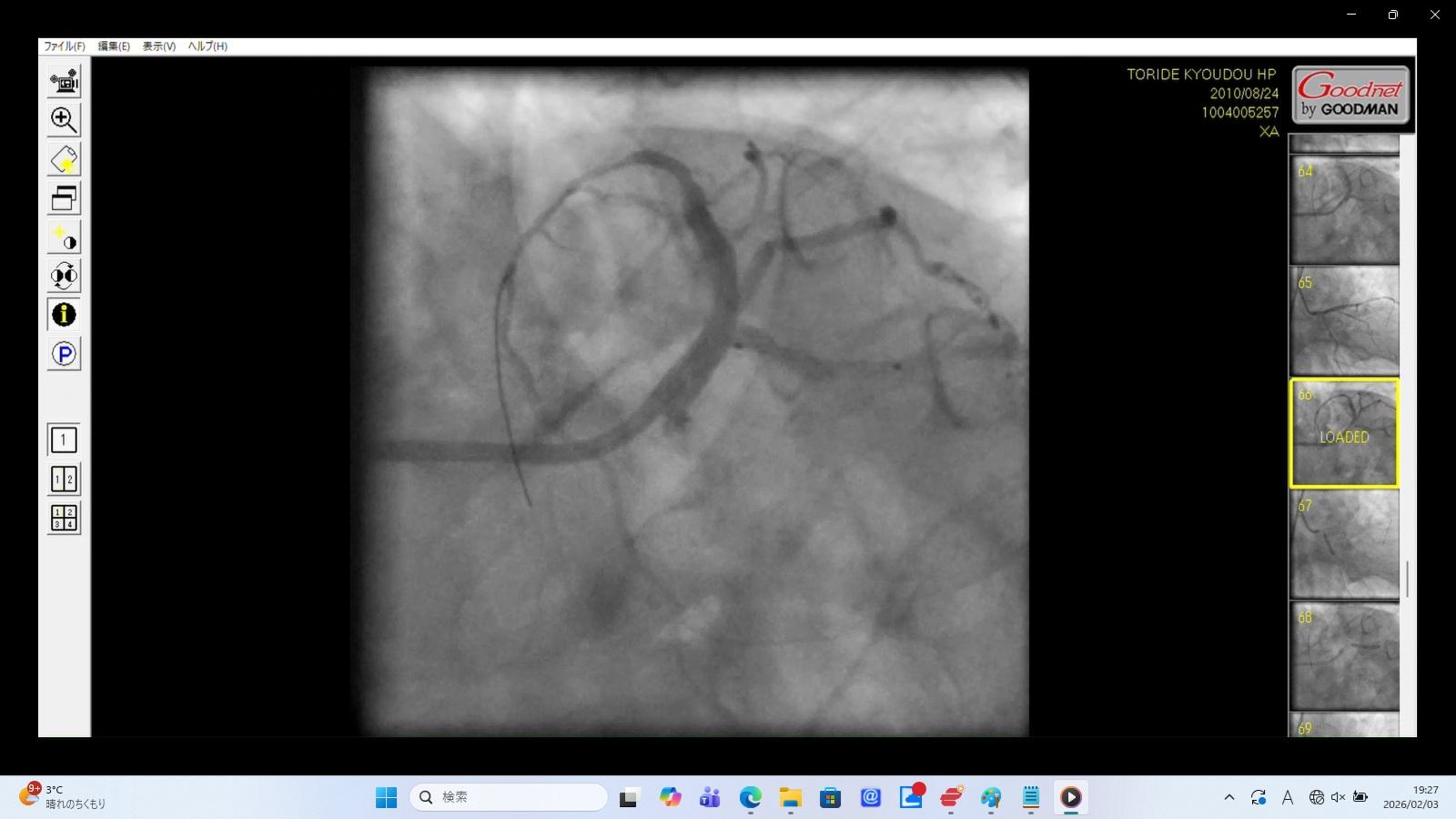
XA0072.mp4:SHA256:XXX
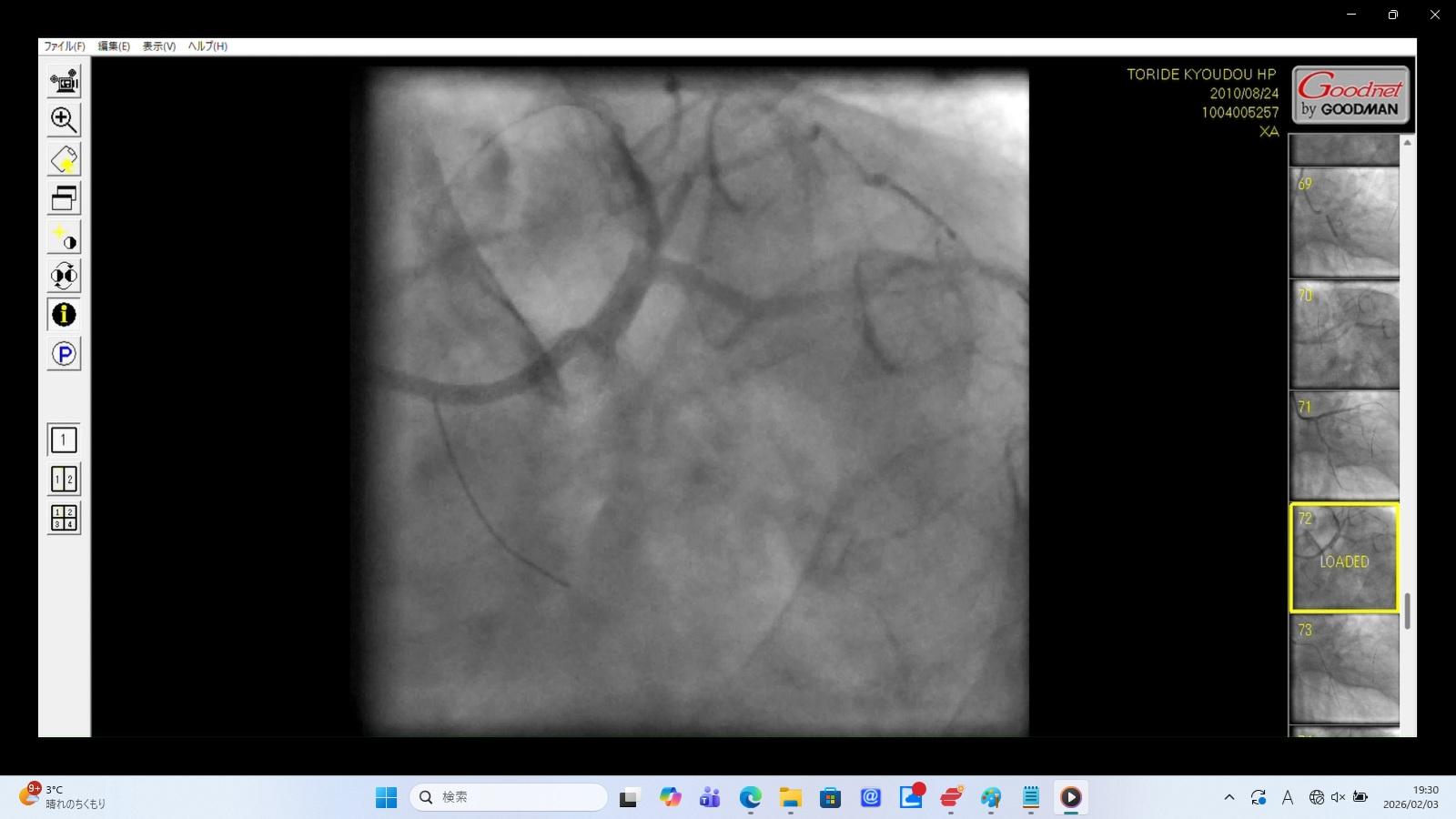
Medical Findings:
Guidewire Position:
The distal guidewire tip is visualized outside the expected anatomical boundaries of the LAD lumen.
Contrast Extravasation:
Accumulation of contrast media is visible in the pericardial space, producing a staining pattern consistent with leakage beyond the vessel wall.
Interpretive Note:
These findings are compatible with a Type III coronary perforation, a complication known to cause rapid hemodynamic deterioration and often requiring urgent pericardiocentesis or surgical intervention.
5. Diffuse Contrast Leakage (XA0019.mp4)
XA0019.mp4:SHA256:XXX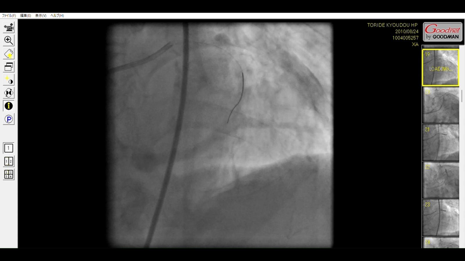
Finding:
Pulsatile, cloud‑like movement of contrast media is observed in the lower‑left quadrant of the frame, occurring independently of injection timing.
Interpretive Note:
This pattern is consistent with contrast and blood entering the pericardial space, a finding typically associated with evolving cardiac tamponade.
6. Left Main Trunk (LMT) Flow Interruption (XA0082.mp4)
XA0082.mp4:SHA256:XXX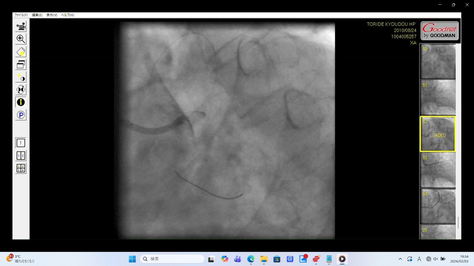
Medical Findings:
Contrast Reflux:
Upon injection, contrast fails to enter the coronary ostium and instead refluxes into the aortic root.
Flow Interruption:
The abrupt cessation of forward flow at the LMT ostium is suggestive of significant structural compromise, such as dissection or mechanical obstruction.
Interpretive Note:
Interruption of LMT flow can critically reduce perfusion to a large portion of the myocardium and typically requires immediate clinical intervention.
Review of Missing Procedural Imaging Intervals
Identified Gaps in Available Video Records
• Gap 1 (27 minutes):
No video files exist between the documented procedure start time (22:35) and the first available file (23:02).
This interval corresponds to the period during which the vascular access route was changed.
• Gap 2 (18 minutes):
No video files exist between 23:08 and 23:26.
This timeframe aligns with nursing notes describing “distal occlusion” and “thrombectomy maneuvers.”
Contextual Note:
Subsequent imaging files show significant vascular injury, including LMT dissection and perforation.
The absence of video during the highest‑risk procedural moments highlights a notable discrepancy between the documented clinical events and the available imaging record.
Chain‑of‑Custody Documentation
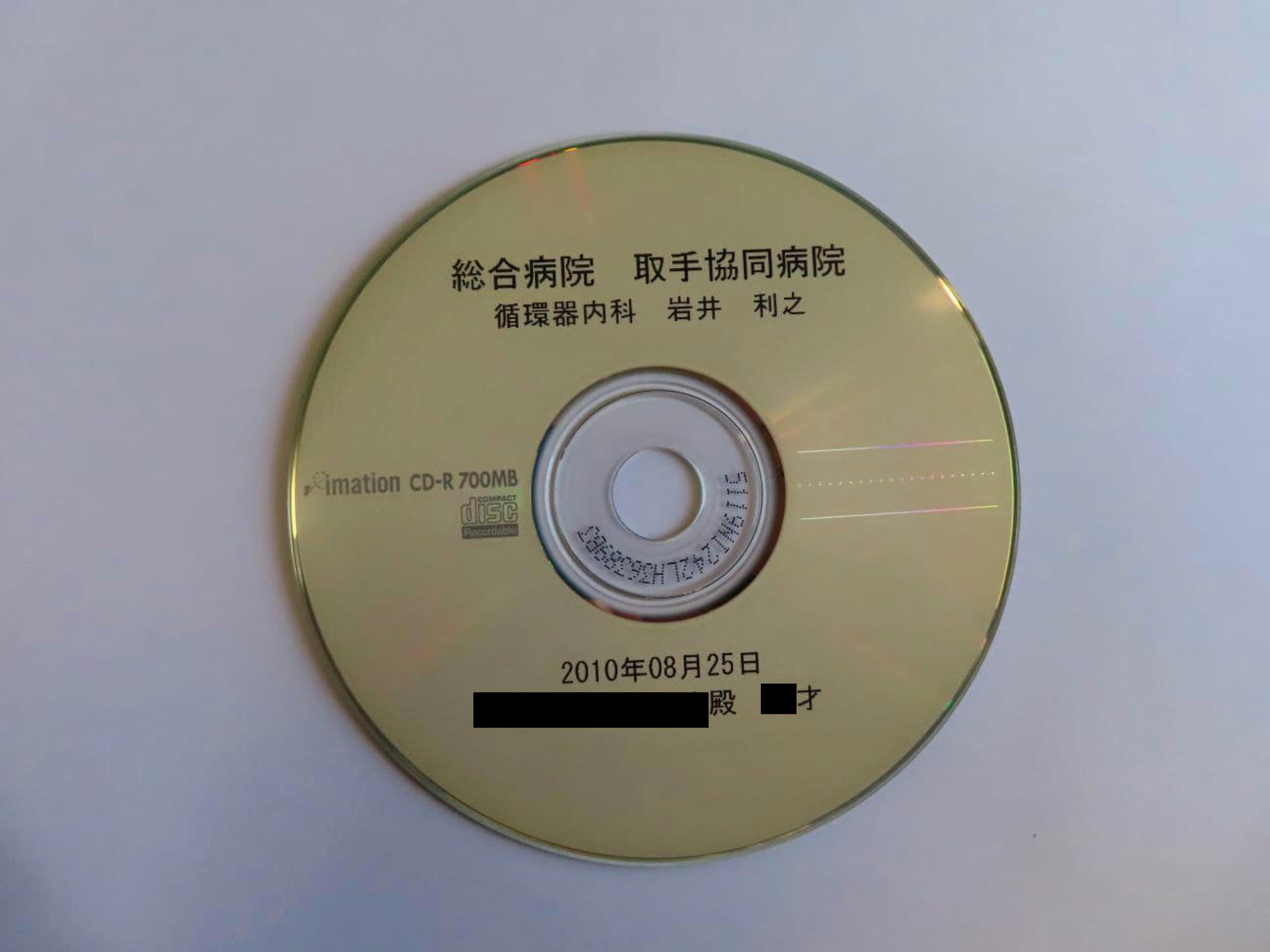
Item:
Photograph of the original CD‑R obtained through the evidence‑preservation process.
Markings:
The disc bears the institutional identifiers of “Toride Kyoudou Hospital” and the “Toride Police Station (Ibaraki Prefecture).”
Contextual Note:
These markings indicate that the digital imaging data were formally transferred to law‑enforcement custody.
The subsequent administrative conclusion that “no findings were present” differs from the content visible in the archived fluoroscopic tracks, representing a significant discrepancy between the recorded imagery and the official assessment.
3. Native Video Track Index (Tracks XA0001 – XA0090)
Researchers, investigative journalists, forensic imaging specialists, and medical peer‑review teams can directly access and examine the native fluoroscopic video files listed below.